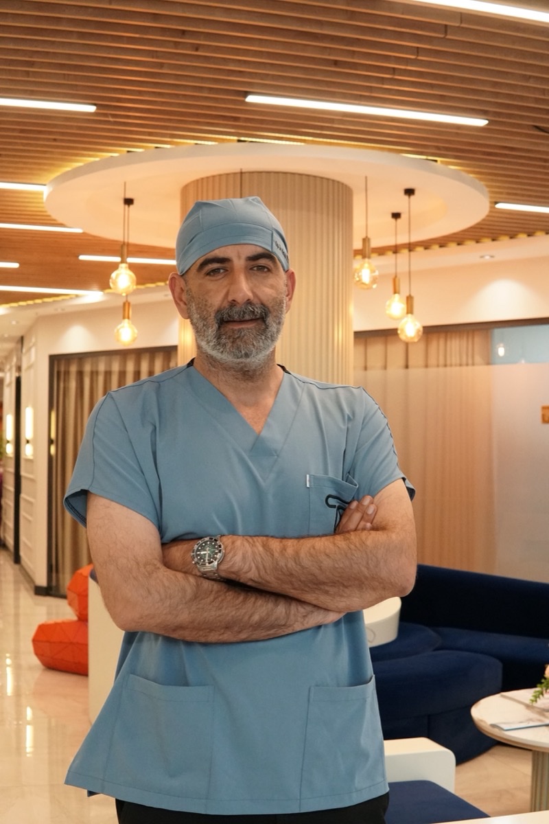
Microsurgical Varicocelectomy and Medical Standards
Varicocele is the most common surgically correctable cause of male infertility, characterized by the pathological dilation of the venous plexus in the scrotum and retrograde blood flow. In modern andrology, the gold standard treatment is microsurgical varicocelectomy. Performed under high-power microscopic magnification, this surgical approach ensures maximum preservation of healthy structures while obliterating diseased veins.
1. Preoperative Evaluation and Preparation
The success of the procedure relies on comprehensive preoperative assessment. Physical examination is conducted in the standing position using the Valsalva maneuver, followed by Scrotal Color Doppler Ultrasound to measure vein diameters and confirm venous reflux. Patient preparation for general or spinal anesthesia is performed under strict sterile surgical protocols.
2. Surgical Incision and Spermatic Cord Exposure
The operation begins with a minimal subinguinal incision of approximately 2 to 3 centimeters. The subinguinal approach avoids transecting abdominal muscle tissues, significantly reducing postoperative pain and facilitating rapid tissue recovery. Dissection proceeds through the fascial layers to expose and isolate the spermatic cord.
3. Microscopic Dissection Under High Magnification
Once the spermatic cord is elevated, an operating microscope providing 8x to 15x magnification is introduced into the surgical field. High magnification is essential to differentiate delicate anatomical structures. Under the microscope, the testicular artery, lymphatic channels, vas deferens, and dilated veins are clearly visualized.
4. Preservation of Testicular Arteries and Lymphatics
The primary advantage of the microsurgical technique over conventional open or laparoscopic approaches is the precise preservation of lymphatic vessels and testicular arteries. Preserving the delicate arteries prevents testicular atrophy, while sparing the clear lymphatic channels prevents postoperative hydrocele formation.
5. Ligation and Division of Pathological Veins
After isolating the vital structures, all internal and external spermatic veins responsible for elevated scrotal temperature and impaired spermatogenesis are individually ligated with fine sutures and divided. Thorough ligation of all venous tributaries reduces the recurrence rate to less than 1-2%.
6. Tissue Closure and Postoperative Care
Following completion of vein ligation, meticulous hemostasis is ensured. The spermatic cord is returned to its anatomical bed, and the surgical layers are closed with absorbable aesthetic sutures. Patients are typically discharged on the same day or within 24 hours. Strenuous physical activity and sexual intercourse should be avoided for 3 to 4 weeks, with semen quality improvement routinely evaluated at 3 and 6 months postoperatively.